Upper Back Back Bones Diagram / Pelvis | Pelvis anatomy, Hip anatomy, Medical anatomy : The third thoracic vertebrae is a small vertebra in the upper middle back that plays an integral role in supporting the rib cage.
byAdmin-
0
Upper Back Back Bones Diagram / Pelvis | Pelvis anatomy, Hip anatomy, Medical anatomy : The third thoracic vertebrae is a small vertebra in the upper middle back that plays an integral role in supporting the rib cage.. The breadth of the back is created by the shoulders at the top and the pelvis at the bottom. The rib cage also anchors the bones of the head, neck, shoulders, and arms to the trunk of the body. Ulna is a long bone. Collectively, these three sections make a tower of 24 bones that gives the body structure and. Bones, discs, and joints in your lower back.
The rib cage also anchors the bones of the head, neck, shoulders, and arms to the trunk of the body. Thoracic region of the spine. The body is the largest part of the vertebrae and the part that bears the most weight. It consists of seven vertebrae. The back's muscles start at the top of the back (named the cervical vertebrae) and go to the tailbone (also named the coccyx).
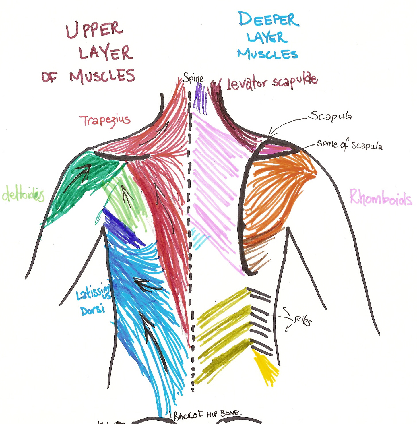
Back Muscles pistures : Biological Science Picture ... from pulpbits.net The breadth of the back is created by the shoulders at the top and the pelvis at the bottom. Human muscles · may 25, 2020. The thoracic spine starts beneath the neck and is comprised of 12 vertebrae, labeled t1 through t12, which go down the back of the torso (figure 1).unlike the cervical spine and lumbar spine, the thoracic spine is relatively immobile because each of its vertebrae are connected to a pair of ribs (one on. Now take your left hand and interlace it around the right arm. There are 12 bones that make up the upper back, which doctors call the thoracic spine. The four principal types of bones are long, short, flat and irregular. The trapezius and latissimus dorsi muscles connect the upper limb to the vertebral column. The back functions are many, such as to house and protect the spinal cord, hold the body and head upright, and adjust the movements of the upper and lower limbs.
However, irritation of the large back and.
Individual anatomical structures include 2: Your spine is made up of 30 bones stacked in a column. It runs from the neck to the upper back. This cancellous bone is in turn, covered by a thin coating of cortical bone (or compact bone), the hard and dense type of osseous tissue. The vertebral column of the lower back includes the five lumbar vertebrae, the sacrum, and the coccyx. Back of right upper extremity. Bones of the pelvis and lower back. Certain back muscles extend to other areas, like the shoulders, upper arms, and thighs. The human back, also called the dorsum, is the large posterior area of the human body, rising from the top of the buttocks to the back of the neck. It is very stiff, and the thoracic spine has a limited range of motion. It runs from the neck to the upper back. There are 12 vertebrae in the thoracic spine. Bone diagram forehead (frontal bone) nose bones (nasals) cheek bone (zygoma) upper jaw (maxilla) lower jaw (mandible) breast bone (sternum) upper arm bone (humerus) lower arm bone (ulna) thigh bone (femur) collar bone (clavicle) toe bones.
Read thoracic spine anatomy and upper back pain. The bones of the chest and upper back combine to form the strong, protective rib cage around the vital thoracic organs such as the heart and lungs. Csm happens when your spinal cord becomes compressed, usually because of degenerative changes in your spine due to age. Both the deltoid and the trapezius are firmly attached to … The rib cage also anchors the bones of the head, neck, shoulders, and arms to the trunk of the body.
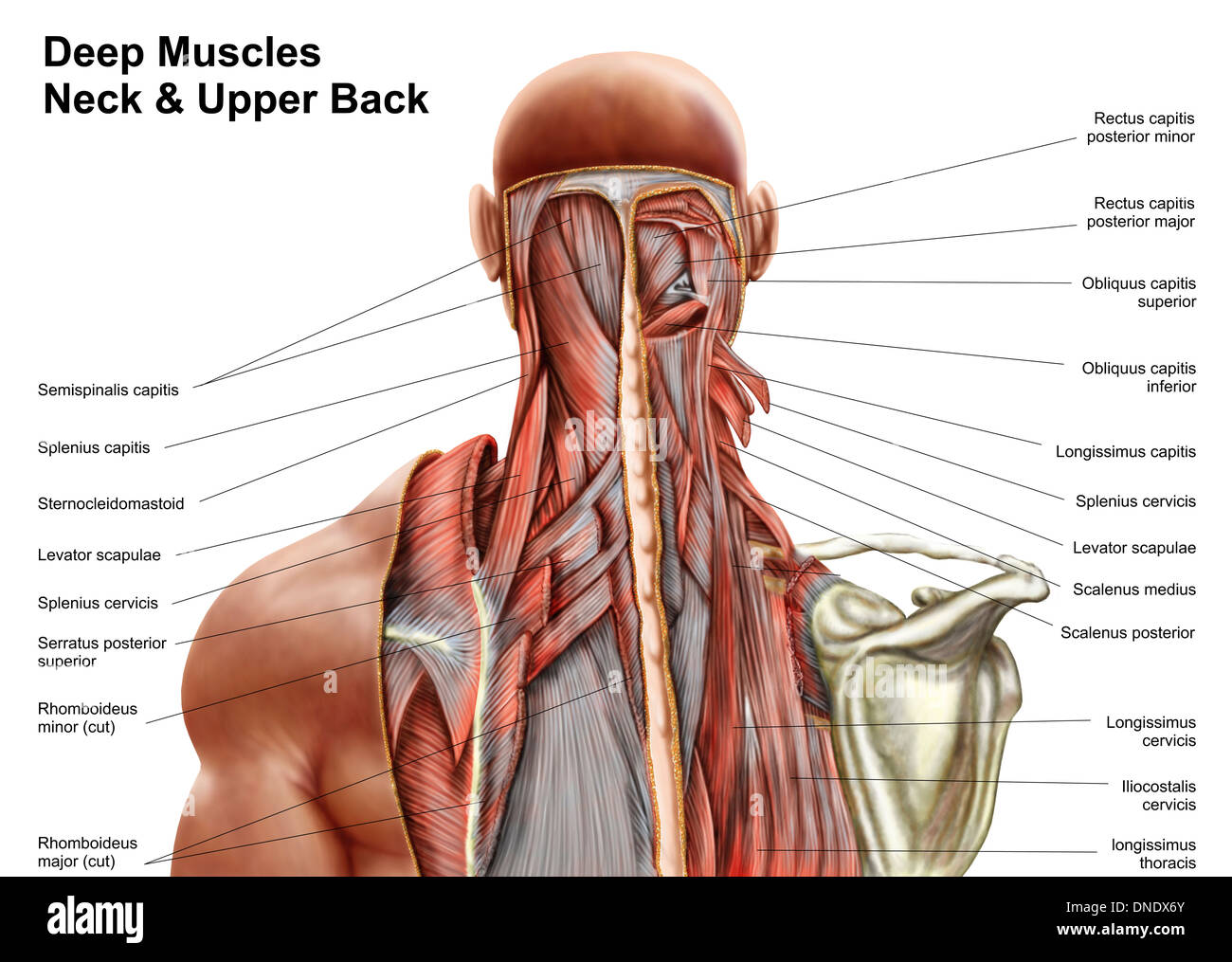
Muscles of the Back - TeachMeAnatomy from s3.amazonaws.com Powerful muscles that move the head and arms attach to these bones as well. The cervical spine protects the nerves connecting to the brain, allowing the head to move freely while supporting its weight. The bones of the chest and upper back combine to form the strong, protective rib cage around the vital thoracic organs such as the heart and lungs. Each vertebra consists of the following parts: The trapezius and latissimus dorsi muscles connect the upper limb to the vertebral column. It runs from the neck to the upper back. This is my video about the muscles of the back. The bones of the chest and upper back combine to form the strong, protective rib cage around the vital thoracic organs such as the heart and lungs.
The rib cage also anchors the bones of the head, neck, shoulders, and arms to the trunk of the body.
Upper back and neck tingling. The human back, also called the dorsum, is the large posterior area of the human body, rising from the top of the buttocks to the back of the neck. Csm happens when your spinal cord becomes compressed, usually because of degenerative changes in your spine due to age. Sometimes you feel the effects right away. The rib cage also anchors the bones of the head, neck, shoulders, and arms to the trunk of the body. Any of these structures can become irritated or inflamed in response to a variety of different factors and conditions, such as poor posture, overuse, trauma, arthritis, and bone cancer.however, most upper back pain causes involve muscle irritation or joint. If you notice a tingling or numbness in your back and also your neck, you may be experiencing a condition called cervical spondylotic myelopathy (csm). The bones of the pelvis and lower back work together to support the body's weight, anchor the abdominal and hip muscles, and protect the delicate vital organs of the vertebral and abdominopelvic cavities. Back of skull (occipital bone) fused vertebrae (5) (sacrum) hand bones (metacarpals) finger bones. The trapezius and latissimus dorsi muscles connect the upper limb to the vertebral column. The pelvis at the bottom of the back and the shoulders at the top of the back give the back its breadth, and it narrows in between these two regions. The third thoracic vertebrae is a small vertebra in the upper middle back that plays an integral role in supporting the rib cage. The lumbar spine connects to the thoracic spine above and the hips below.
Collectively, these three sections make a tower of 24 bones that gives the body structure and. The trapezius and latissimus dorsi muscles connect the upper limb to the vertebral column. License image the deltoid, teres major, teres minor, infraspinatus, supraspinatus (not shown) and subscapularis muscles (not shown) all extend from the scapula to the humerus and act on the shoulder joint. In addition to the thoracic spine and shoulder blades, there are numerous nerves, muscles, tendons, and ligaments in the upper back. Thoracic region of the spine.
Back Muscle Anatomy Model - Human Anatomy from c8.alamy.com From the side, your vertebral column has a natural curve toward the back of your body as it passes through your upper chest, balanced by a similar curve toward the front through the lower vertebrae. Human muscles · may 25, 2020. These bones are connected at the back with specialized joints. Bones of the chest and upper back / the muscles, bones, ligaments, and tendons in the back can all be injured and cause back. The thoracic spine sits between the cervical spine in the neck and the lumbar spine in the lower back. The pelvis at the bottom of the back and the shoulders at the top of the back give the back its breadth, and it narrows in between these two regions. The rib cage also anchors the bones of the head, neck, shoulders, and arms to the trunk of the body. However, irritation of the large back and.
Anatomy upper limb bones and cartilages bones of upper limb.
The back functions are many, such as to house and protect the spinal cord, hold the body and head upright, and adjust the movements of the upper and lower limbs. Bones, discs, and joints in your lower back. Back of skull (occipital bone) fused vertebrae (5) (sacrum) hand bones (metacarpals) finger bones. Csm happens when your spinal cord becomes compressed, usually because of degenerative changes in your spine due to age. The muscles of the lower back help stabilize, rotate, flex, and extend the spinal column, which is a bony tower of 24 vertebrae that gives the body structure and houses the spinal cord. Your spine is made up of 30 bones stacked in a column. Bone diagram back skeletal dysplasias affect the development and growth of cartilage bones and joints causing abnormally shaped bones especially in the head spine and f i g u r e 1 diagram of. Certain back muscles extend to other areas, like the shoulders, upper arms, and thighs. The human back, also called the dorsum, is the large posterior area of the human body, rising from the top of the buttocks to the back of the neck. From the side, your vertebral column has a natural curve toward the back of your body as it passes through your upper chest, balanced by a similar curve toward the front through the lower vertebrae. The breadth of the back is created by the shoulders at the top and the pelvis at the bottom. The cervical spine protects the nerves connecting to the brain, allowing the head to move freely while supporting its weight. 12 photos of the human back bone chart.
Place right elbow on left elbow back bones diagram. It consists of seven vertebrae.